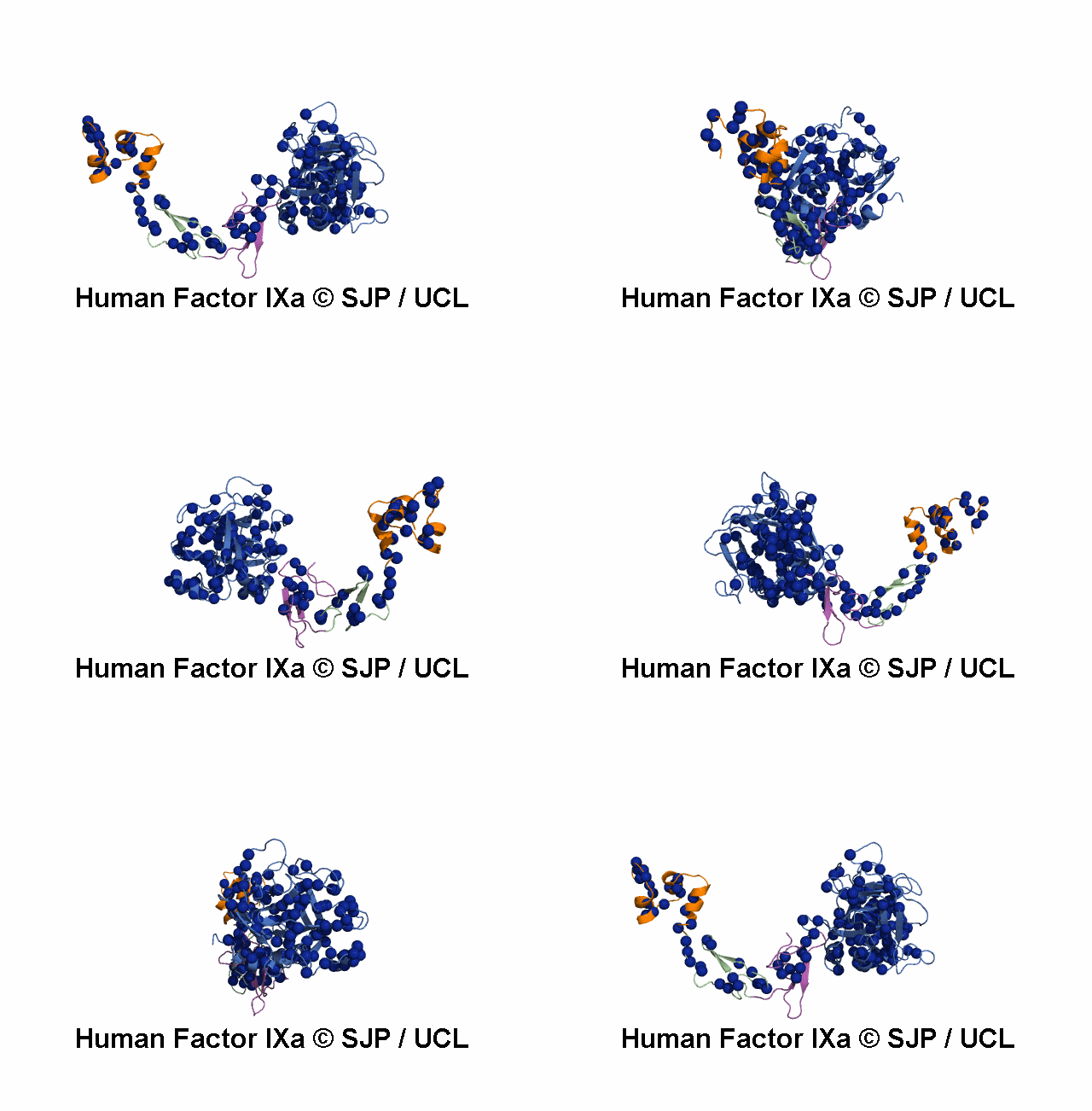
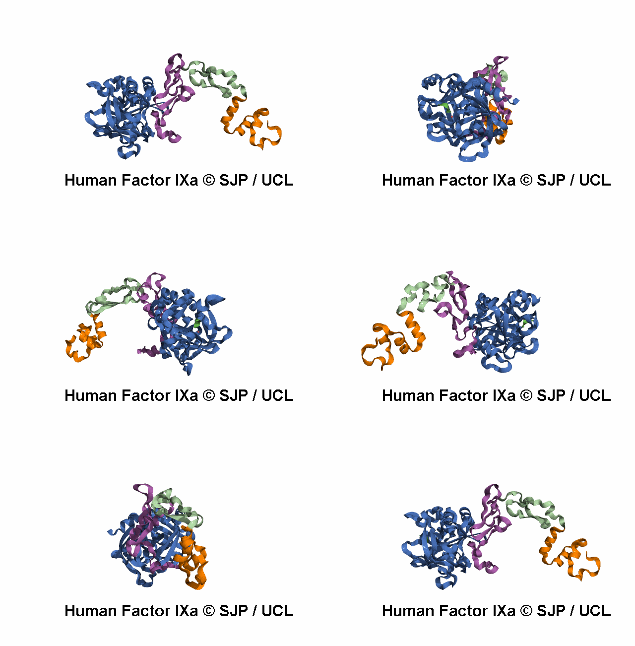
PDB |
Method |
Description |
Resolution |
Chain |
Position |
PDBsum |
|
1CFH |
NMR |
structure of the metal-free gamma-carboxyglutamic acid-rich membrane binding region of factor ix by two-dimensional nmr spectroscopy |
- |
A |
47-93 |
>>
|
|
1CFI |
NMR |
nmr structure of calcium ion-bound gamma-carboxy-glutamic acid-rich domain of factor ix |
- |
A |
47-93 |
>>
|
|
1EDM |
X-ray |
epidermal growth factor-like domain from human factor ix |
1.50 |
B/C |
92-130 |
>>
|
|
1IXA |
NMR |
the three-dimensional structure of the first egf-like module of human factor ix: comparison with egf and tgf-a |
- |
A |
92-130 |
>>
|
|
1MGX |
NMR |
coagulation factor, mg(ii), nmr, 7 structures (backbone atoms only) |
- |
A |
47-93 |
>>
|
|
1NL0 |
X-ray |
crystal structure of human factor ix gla domain in complex of an inhibitory antibody, 10c12 |
2.20 |
G |
47-91 |
>>
|
|
1RFN |
X-ray |
human coagulation factor ixa in complex with p-amino benzamidine |
2.80 |
A |
227-461 |
>>
|
|
1RFN |
X-ray |
human coagulation factor ixa in complex with p-amino benzamidine |
2.80 |
B |
133-188 |
>>
|
|
2WPH |
X-ray |
factor ixa superactive triple mutant |
1.50 |
E |
133-191 |
>>
|
|
2WPH |
X-ray |
factor ixa superactive triple mutant |
1.50 |
S |
227-461 |
>>
|
|
2WPI |
X-ray |
factor ixa superactive double mutant |
1.99 |
E |
133-191 |
>>
|
|
2WPI |
X-ray |
factor ixa superactive double mutant |
1.99 |
S |
227-461 |
>>
|
|
2WPJ |
X-ray |
factor ixa superactive triple mutant, nacl-soaked |
1.60 |
E |
133-191 |
>>
|
|
2WPJ |
X-ray |
factor ixa superactive triple mutant, nacl-soaked |
1.60 |
S |
227-461 |
>>
|
|
2WPK |
X-ray |
factor ixa superactive triple mutant, ethylene glycol-soaked |
2.21 |
E |
133-191 |
>>
|
|
2WPK |
X-ray |
factor ixa superactive triple mutant, ethylene glycol-soaked |
2.21 |
S |
227-461 |
>>
|
|
2WPL |
X-ray |
factor ixa superactive triple mutant, edta-soaked |
1.82 |
E |
133-191 |
>>
|
|
2WPL |
X-ray |
factor ixa superactive triple mutant, edta-soaked |
1.82 |
S |
227-461 |
>>
|
|
2WPM |
X-ray |
factor ixa superactive mutant, egr-cmk inhibited |
2.00 |
E |
133-191 |
>>
|
|
2WPM |
X-ray |
factor ixa superactive mutant, egr-cmk inhibited |
2.00 |
S |
227-461 |
>>
|
|
3KCG |
X-ray |
crystal structure of the antithrombin-factor ixa-pentasaccharide complex |
1.70 |
H |
227-461 |
>>
|
|
3KCG |
X-ray |
crystal structure of the antithrombin-factor ixa-pentasaccharide complex |
1.70 |
L |
131-188 |
>>
|
|
3LC3 |
X-ray |
benzothiophene inhibitors of factor ixa |
1.90 |
A/C |
227-461 |
>>
|
|
3LC3 |
X-ray |
benzothiophene inhibitors of factor ixa |
1.90 |
B/D |
133-188 |
>>
|
|
3LC5 |
X-ray |
selective benzothiophine inhibitors of factor ixa |
2.62 |
A |
227-461 |
>>
|
|
3LC5 |
X-ray |
selective benzothiophine inhibitors of factor ixa |
2.62 |
B |
133-188 |
>>
|
|
4WM0 |
X-ray |
crystal structure of mouse xyloside xylosyltransferase 1 complexed with acceptor ligand |
2.37 |
D |
92-130 |
>>
|
|
4WMA |
X-ray |
crystal structure of mouse xyloside xylosyltransferase 1 complexed with manganese,acceptor ligand and udp-glucose |
1.62 |
D |
92-130 |
>>
|
|
4WMB |
X-ray |
crystal structure of mouse xyloside xylosyltransferase 1 complexed with manganese, acceptor ligand and udp |
2.05 |
D |
92-130 |
>>
|
|
4WMI |
X-ray |
crystal structure of mouse xyloside xylosyltransferase 1 complexed with manganese, product ligand and udp (product complex i) |
1.87 |
D |
92-130 |
>>
|
|
4WMK |
X-ray |
crystal structure of mouse xyloside xylosyltransferase 1 complexed with manganese, product ligand and udp (product complex ii) |
2.08 |
D |
92-130 |
>>
|
|
4WN2 |
X-ray |
crystal structure of mouse xyloside xylosyltransferase 1 complexed with manganese, product ligand and udp (product complex iii) |
1.95 |
D |
92-130 |
>>
|
|
4WNH |
X-ray |
crystal structure of mouse xyloside xylosyltransferase 1 complexed with manganese,acceptor ligand and udp-xylose |
1.95 |
D |
92-130 |
>>
|
|
4YZU |
X-ray |
rapid development of two factor ixa inhibitors from hit to lead |
1.41 |
A |
227-461 |
>>
|
|
4YZU |
X-ray |
rapid development of two factor ixa inhibitors from hit to lead |
1.41 |
B |
130-191 |
>>
|
|
4Z0K |
X-ray |
rapid development of two factor ixa inhibitors from hit to lead |
1.41 |
A |
227-461 |
>>
|
|
4Z0K |
X-ray |
rapid development of two factor ixa inhibitors from hit to lead |
1.41 |
B |
130-191 |
>>
|
|
4ZAE |
X-ray |
development of a novel class of potent and selective fixa inhibitors |
1.86 |
A |
227-461 |
>>
|
|
4ZAE |
X-ray |
development of a novel class of potent and selective fixa inhibitors |
1.86 |
B |
130-191 |
>>
|
|
5EGM |
X-ray |
development of a novel tricyclic class of potent and selective fixa inhibitors |
1.84 |
A |
227-461 |
>>
|
|
5EGM |
X-ray |
development of a novel tricyclic class of potent and selective fixa inhibitors |
1.84 |
B |
130-191 |
>>
|
|
5F84 |
X-ray |
crystal structure of drosophila poglut1 (rumi) complexed with its glycoprotein product (glucosylated egf repeat) and udp |
2.50 |
A |
21-407 |
>>
|
|
5F84 |
X-ray |
crystal structure of drosophila poglut1 (rumi) complexed with its glycoprotein product (glucosylated egf repeat) and udp |
2.50 |
B |
130-191 |
>>
|
|
5F85 |
X-ray |
crystal structure of drosophila poglut1 (rumi) complexed with its substrate protein (egf repeat) and udp |
2.15 |
A |
21-407 |
>>
|
|
5F85 |
X-ray |
crystal structure of drosophila poglut1 (rumi) complexed with its substrate protein (egf repeat) and udp |
2.15 |
B |
92-130 |
>>
|
|
5F86 |
X-ray |
crystal structure of drosophila poglut1 (rumi) complexed with its substrate protein (egf repeat) |
1.90 |
A |
21-407 |
>>
|
|
5F86 |
X-ray |
crystal structure of drosophila poglut1 (rumi) complexed with its substrate protein (egf repeat) |
1.90 |
B |
92-130 |
>>
|
|
5JB8 |
X-ray |
crystal structure of factor ixa variant k98t in complex with egr-chloromethylketone |
1.45 |
E |
134-191 |
>>
|
|
5JB8 |
X-ray |
crystal structure of factor ixa variant k98t in complex with egr-chloromethylketone |
1.45 |
S |
227-461 |
>>
|
|
5JB9 |
X-ray |
crystal structure of factor ixa k98t variant in complex with ppack |
1.30 |
E |
134-191 |
>>
|
|
5JB9 |
X-ray |
crystal structure of factor ixa k98t variant in complex with ppack |
1.30 |
S |
227-461 |
>>
|
|
5JBA |
X-ray |
crystal structure of factor ixa variant v16i k98t y177t i212v in complex with ppack |
1.40 |
E |
134-191 |
>>
|
|
5JBA |
X-ray |
crystal structure of factor ixa variant v16i k98t y177t i212v in complex with ppack |
1.40 |
S |
227-461 |
>>
|
|
5JBB |
X-ray |
crystal structure of factor ixa variant v16i k98t y177t i213v in complex with egr-chloromethylketone |
1.56 |
E |
134-191 |
>>
|
|
5JBB |
X-ray |
crystal structure of factor ixa variant v16i k98t y177t i213v in complex with egr-chloromethylketone |
1.56 |
S |
227-461 |
>>
|
|
5JBC |
X-ray |
crystal structure of factor ixa variant v16i k98t y177t i213v in complex with ppack |
1.90 |
E |
134-191 |
>>
|
|
5JBC |
X-ray |
crystal structure of factor ixa variant v16i k98t y177t i213v in complex with ppack |
1.90 |
S |
227-461 |
>>
|
|
5TNO |
X-ray |
discovery of novel aminobenzisoxazole derivatives as orally available factor ixa inhibitors |
1.54 |
A |
227-461 |
>>
|
|
5TNO |
X-ray |
discovery of novel aminobenzisoxazole derivatives as orally available factor ixa inhibitors |
1.54 |
B |
130-191 |
>>
|
|
5TNT |
X-ray |
discovery of novel aminobenzisoxazole derivatives as orally available factor ixa inhibitors |
1.40 |
A |
227-461 |
>>
|
|
5TNT |
X-ray |
discovery of novel aminobenzisoxazole derivatives as orally available factor ixa inhibitors |
1.40 |
B |
130-191 |
>>
|
|
5VYG |
X-ray |
crystal structure of hfa9 egf repeat with o-glucose trisaccharide |
2.20 |
A/B/C |
92-130 |
>>
|
|
6MV4 |
X-ray |
crystal structure of human coagulation factor ixa |
1.37 |
H |
227-461 |
>>
|
|
6MV4 |
X-ray |
crystal structure of human coagulation factor ixa |
1.37 |
L |
132-185 |
>>
|
|
6RFK |
X-ray |
crystal structure of egrck-inhibited gla-domainless fixa (k148q, r150q variant) |
1.60 |
E |
130-191 |
>>
|
|
6RFK |
X-ray |
crystal structure of egrck-inhibited gla-domainless fixa (k148q, r150q variant) |
1.60 |
S |
227-461 |
>>
|
|
6X5J |
X-ray |
discovery of hydroxy pyrimidine factor ixa inhibitors |
2.51 |
B |
130-191 |
>>
|
|
6X5J |
X-ray |
discovery of hydroxy pyrimidine factor ixa inhibitors |
2.51 |
A |
227-461 |
>>
|
|
6X5L |
X-ray |
discovery of hydroxy pyrimidine factor ixa inhibitors |
2.25 |
A |
227-460 |
>>
|
|
6X5L |
X-ray |
discovery of hydroxy pyrimidine factor ixa inhibitors |
2.25 |
B |
130-191 |
>>
|
|
6X5P |
X-ray |
discovery of hydroxy pyrimidine factor ixa inhibitors |
2.00 |
A |
227-461 |
>>
|
|
6X5P |
X-ray |
discovery of hydroxy pyrimidine factor ixa inhibitors |
2.00 |
B |
130-191 |
>>
|
|
NA |
prediction |
alphafold structure prediction for coagulation factor ix. |
NA |
A |
1-461 |
>>
|


